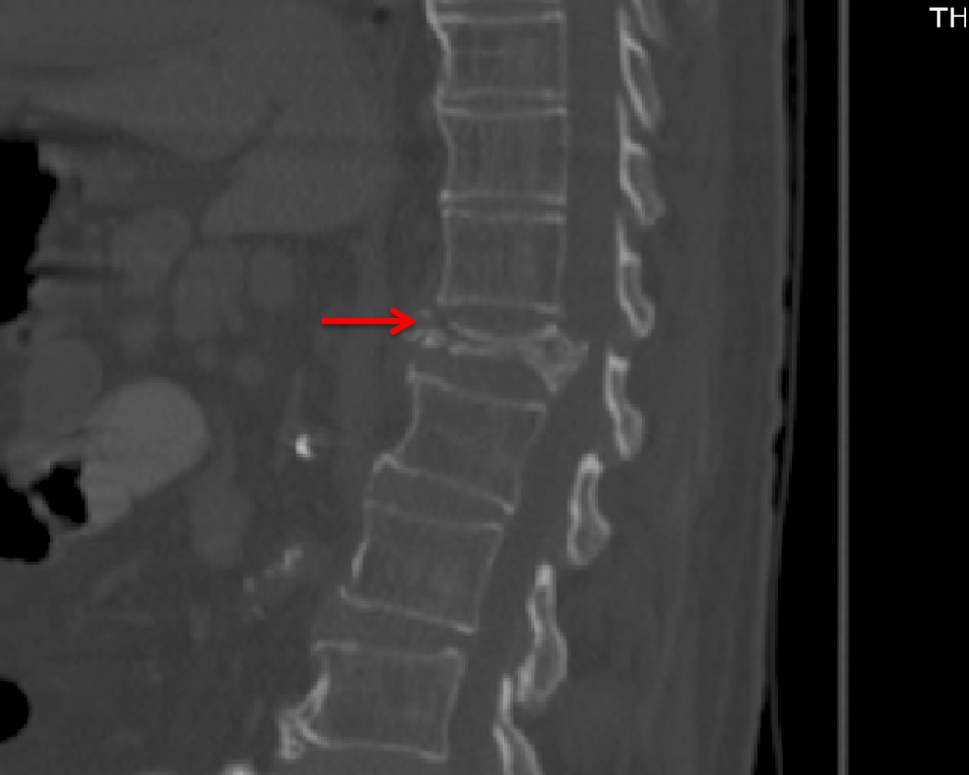
Compression Fracture With Retropulsion Radiology . Retropulsion of a fragment is the typical feature of a burst fracture and distinguishes it clearly from. in special cases where the compression fracture is because of an infectious or malignant process, more advanced mri. In the absence of these signs fractures are. the ideal patient has clinical and imaging evidence of a vcf less than 3 weeks old and is refractory to nonoperative. though exceedingly rare, occasionally retropulsion of fracture fragments may result in compression of the spinal cord. Preserved normal fatty bone marrow. retropulsion of posterosuperior vertebral body fragment. on conventional imaging, acute fracture signs include cortical breaking or impaction of trabeculae; However, in some cases this pain is only mild and attributed to another issue like a muscle spasm. the following features favor the diagnosis of a benign compression fracture:
from exomzwfis.blob.core.windows.net
the following features favor the diagnosis of a benign compression fracture: on conventional imaging, acute fracture signs include cortical breaking or impaction of trabeculae; in special cases where the compression fracture is because of an infectious or malignant process, more advanced mri. the ideal patient has clinical and imaging evidence of a vcf less than 3 weeks old and is refractory to nonoperative. In the absence of these signs fractures are. retropulsion of posterosuperior vertebral body fragment. Retropulsion of a fragment is the typical feature of a burst fracture and distinguishes it clearly from. However, in some cases this pain is only mild and attributed to another issue like a muscle spasm. though exceedingly rare, occasionally retropulsion of fracture fragments may result in compression of the spinal cord. Preserved normal fatty bone marrow.
What Is Compression Fracture With Retropulsion at Deborah Noel blog
Compression Fracture With Retropulsion Radiology retropulsion of posterosuperior vertebral body fragment. Preserved normal fatty bone marrow. retropulsion of posterosuperior vertebral body fragment. However, in some cases this pain is only mild and attributed to another issue like a muscle spasm. the ideal patient has clinical and imaging evidence of a vcf less than 3 weeks old and is refractory to nonoperative. though exceedingly rare, occasionally retropulsion of fracture fragments may result in compression of the spinal cord. Retropulsion of a fragment is the typical feature of a burst fracture and distinguishes it clearly from. on conventional imaging, acute fracture signs include cortical breaking or impaction of trabeculae; In the absence of these signs fractures are. the following features favor the diagnosis of a benign compression fracture: in special cases where the compression fracture is because of an infectious or malignant process, more advanced mri.
From animalia-life.club
Compression Fracture Diagram Compression Fracture With Retropulsion Radiology Retropulsion of a fragment is the typical feature of a burst fracture and distinguishes it clearly from. on conventional imaging, acute fracture signs include cortical breaking or impaction of trabeculae; in special cases where the compression fracture is because of an infectious or malignant process, more advanced mri. However, in some cases this pain is only mild and. Compression Fracture With Retropulsion Radiology.
From jpay-tj.blogspot.com
L1 Compression Fracture / What Is An L1 Compression Fracture A Closer Compression Fracture With Retropulsion Radiology on conventional imaging, acute fracture signs include cortical breaking or impaction of trabeculae; Retropulsion of a fragment is the typical feature of a burst fracture and distinguishes it clearly from. Preserved normal fatty bone marrow. though exceedingly rare, occasionally retropulsion of fracture fragments may result in compression of the spinal cord. in special cases where the compression. Compression Fracture With Retropulsion Radiology.
From exomzwfis.blob.core.windows.net
What Is Compression Fracture With Retropulsion at Deborah Noel blog Compression Fracture With Retropulsion Radiology the following features favor the diagnosis of a benign compression fracture: the ideal patient has clinical and imaging evidence of a vcf less than 3 weeks old and is refractory to nonoperative. though exceedingly rare, occasionally retropulsion of fracture fragments may result in compression of the spinal cord. retropulsion of posterosuperior vertebral body fragment. Preserved normal. Compression Fracture With Retropulsion Radiology.
From exomzwfis.blob.core.windows.net
What Is Compression Fracture With Retropulsion at Deborah Noel blog Compression Fracture With Retropulsion Radiology on conventional imaging, acute fracture signs include cortical breaking or impaction of trabeculae; Retropulsion of a fragment is the typical feature of a burst fracture and distinguishes it clearly from. Preserved normal fatty bone marrow. However, in some cases this pain is only mild and attributed to another issue like a muscle spasm. the following features favor the. Compression Fracture With Retropulsion Radiology.
From exomzwfis.blob.core.windows.net
What Is Compression Fracture With Retropulsion at Deborah Noel blog Compression Fracture With Retropulsion Radiology Preserved normal fatty bone marrow. Retropulsion of a fragment is the typical feature of a burst fracture and distinguishes it clearly from. the following features favor the diagnosis of a benign compression fracture: retropulsion of posterosuperior vertebral body fragment. in special cases where the compression fracture is because of an infectious or malignant process, more advanced mri.. Compression Fracture With Retropulsion Radiology.
From www.researchgate.net
A spine MRI without contrast revealed a compression fracture of the T8 Compression Fracture With Retropulsion Radiology on conventional imaging, acute fracture signs include cortical breaking or impaction of trabeculae; the ideal patient has clinical and imaging evidence of a vcf less than 3 weeks old and is refractory to nonoperative. retropulsion of posterosuperior vertebral body fragment. Retropulsion of a fragment is the typical feature of a burst fracture and distinguishes it clearly from.. Compression Fracture With Retropulsion Radiology.
From www.researchgate.net
65yearold man with malignant vertebral body compression fracture due Compression Fracture With Retropulsion Radiology However, in some cases this pain is only mild and attributed to another issue like a muscle spasm. retropulsion of posterosuperior vertebral body fragment. the following features favor the diagnosis of a benign compression fracture: Retropulsion of a fragment is the typical feature of a burst fracture and distinguishes it clearly from. Preserved normal fatty bone marrow. . Compression Fracture With Retropulsion Radiology.
From medicaldialogues.in
Radiomics model distinguishes between acute and chronic vertebral Compression Fracture With Retropulsion Radiology the ideal patient has clinical and imaging evidence of a vcf less than 3 weeks old and is refractory to nonoperative. though exceedingly rare, occasionally retropulsion of fracture fragments may result in compression of the spinal cord. retropulsion of posterosuperior vertebral body fragment. Preserved normal fatty bone marrow. in special cases where the compression fracture is. Compression Fracture With Retropulsion Radiology.
From www.ajronline.org
Morphologic Change in Vertebral Body After Percutaneous Vertebroplasty Compression Fracture With Retropulsion Radiology However, in some cases this pain is only mild and attributed to another issue like a muscle spasm. the following features favor the diagnosis of a benign compression fracture: on conventional imaging, acute fracture signs include cortical breaking or impaction of trabeculae; Retropulsion of a fragment is the typical feature of a burst fracture and distinguishes it clearly. Compression Fracture With Retropulsion Radiology.
From www.researchgate.net
Xray images of vertebral compression fracture a) xray images of Compression Fracture With Retropulsion Radiology In the absence of these signs fractures are. in special cases where the compression fracture is because of an infectious or malignant process, more advanced mri. the ideal patient has clinical and imaging evidence of a vcf less than 3 weeks old and is refractory to nonoperative. retropulsion of posterosuperior vertebral body fragment. However, in some cases. Compression Fracture With Retropulsion Radiology.
From in.pinterest.com
Acute and chronic vertebral compression fractures Radiology Case Compression Fracture With Retropulsion Radiology on conventional imaging, acute fracture signs include cortical breaking or impaction of trabeculae; retropulsion of posterosuperior vertebral body fragment. the ideal patient has clinical and imaging evidence of a vcf less than 3 weeks old and is refractory to nonoperative. Retropulsion of a fragment is the typical feature of a burst fracture and distinguishes it clearly from.. Compression Fracture With Retropulsion Radiology.
From radiologykey.com
The Lumbar and Thoracic Spine Radiology Key Compression Fracture With Retropulsion Radiology on conventional imaging, acute fracture signs include cortical breaking or impaction of trabeculae; though exceedingly rare, occasionally retropulsion of fracture fragments may result in compression of the spinal cord. in special cases where the compression fracture is because of an infectious or malignant process, more advanced mri. the ideal patient has clinical and imaging evidence of. Compression Fracture With Retropulsion Radiology.
From mungfali.com
Lumbar Compression Fracture X Ray Compression Fracture With Retropulsion Radiology retropulsion of posterosuperior vertebral body fragment. the following features favor the diagnosis of a benign compression fracture: though exceedingly rare, occasionally retropulsion of fracture fragments may result in compression of the spinal cord. Retropulsion of a fragment is the typical feature of a burst fracture and distinguishes it clearly from. on conventional imaging, acute fracture signs. Compression Fracture With Retropulsion Radiology.
From radiologyassistant.nl
The Radiology Assistant TLICS Classification of fractures Compression Fracture With Retropulsion Radiology the ideal patient has clinical and imaging evidence of a vcf less than 3 weeks old and is refractory to nonoperative. In the absence of these signs fractures are. retropulsion of posterosuperior vertebral body fragment. though exceedingly rare, occasionally retropulsion of fracture fragments may result in compression of the spinal cord. However, in some cases this pain. Compression Fracture With Retropulsion Radiology.
From www.ajnr.org
Review of the Imaging Features of Benign Osteoporotic and Malignant Compression Fracture With Retropulsion Radiology retropulsion of posterosuperior vertebral body fragment. Preserved normal fatty bone marrow. in special cases where the compression fracture is because of an infectious or malignant process, more advanced mri. In the absence of these signs fractures are. However, in some cases this pain is only mild and attributed to another issue like a muscle spasm. though exceedingly. Compression Fracture With Retropulsion Radiology.
From medicine.utah.edu
Lumbar Spine Trauma Radiology U of U School of Medicine Compression Fracture With Retropulsion Radiology retropulsion of posterosuperior vertebral body fragment. the following features favor the diagnosis of a benign compression fracture: in special cases where the compression fracture is because of an infectious or malignant process, more advanced mri. Preserved normal fatty bone marrow. on conventional imaging, acute fracture signs include cortical breaking or impaction of trabeculae; Retropulsion of a. Compression Fracture With Retropulsion Radiology.
From janene-howzell.blogspot.com
compression fracture from car accident janenehowzell Compression Fracture With Retropulsion Radiology In the absence of these signs fractures are. retropulsion of posterosuperior vertebral body fragment. the following features favor the diagnosis of a benign compression fracture: though exceedingly rare, occasionally retropulsion of fracture fragments may result in compression of the spinal cord. the ideal patient has clinical and imaging evidence of a vcf less than 3 weeks. Compression Fracture With Retropulsion Radiology.
From www.medscape.com
Figures Compression Fracture With Retropulsion Radiology In the absence of these signs fractures are. Retropulsion of a fragment is the typical feature of a burst fracture and distinguishes it clearly from. on conventional imaging, acute fracture signs include cortical breaking or impaction of trabeculae; the ideal patient has clinical and imaging evidence of a vcf less than 3 weeks old and is refractory to. Compression Fracture With Retropulsion Radiology.